Leg Bones Diagram / Home Anatomy Physiology For Ems Libguides At Com Library - The stifle joint connects the femur, which is the dog thigh bone, to the tibia and fibula, the lower leg bones, and the patella,the canine equivalent to the knee cap.
Leg Bones Diagram / Home Anatomy Physiology For Ems Libguides At Com Library - The stifle joint connects the femur, which is the dog thigh bone, to the tibia and fibula, the lower leg bones, and the patella,the canine equivalent to the knee cap.. Posted on june 4, 2014 by admin. The authors explore how digitizing one of the seven basic quality tools—the fishbone diagram—using mind mapping can significantly improve the tool. This short post will try to cover the dog leg anatomy in detail with labeled diagrams. The rounded, proximal end is the head of the femur, which articulates with the acetabulum of the hip bone to form the hip joint. Human leg bone diagram :
I think this was the case this is a detailed diagram of a horse's hoof. Also called the shin bone, the tibia is the longer of the two bones in the. The lumbar plexus forms in the lower back from the merger of spinal nerves l1 through l4 while the. The foot bones shown in this diagram are the talus, navicular, cuneiform, cuboid, metatarsals and calcaneus. The bones of the leg and foot form part of the appendicular skeleton that supports the many muscles of the lower limbs.
Bones Of The Leg And The Foot Skeleton Of The Hindlimb Anatomy Americanhighschool Anatomylessons Ahsrocks A Anatomy Bones Leg Anatomy Human Body Anatomy from i.pinimg.com Posted on june 4, 2014 by admin. Cancellous bone produces red blood cells, platelets, and white blood cells. The rounded, proximal end is the head of the femur, which articulates with the acetabulum of the hip bone to form the hip joint. The femur, or thighbone, is the longest and largest bone in the human body. This area is commonly referred to as the calf. The bones of the leg and foot form part of the appendicular skeleton that supports the many muscles of the lower limbs. The authors explore how digitizing one of the seven basic quality tools—the fishbone diagram—using mind mapping can significantly improve the tool. Related posts of diagram of leg bones inside of arm muscle and bone.
Leg bones diagram diagram schematic ideas lower leg muscle diagram blank sketch coloring page antique 1890s medical anatomy diagram leg bones skeleton posted on april 18, 2019april 18, 2019.
Most of the leg skeleton has bony prominences and margins that can be palpated and some serve as anatomical landmarks that define the extent of the leg. Its decrease finish helps create the knee joint. Its lower end helps create the knee joint. Skeletal system diagrams | skeletal system anatomy, human anatomy and physiology.diagram of blood and nerve supply to bone. I think this was the case this is a detailed diagram of a horse's hoof. Posted on june 4, 2014 by admin. The elbow is located below the chest at the back of the foreleg. Related posts of diagram of leg bones inside of arm muscle and bone. Another bone that is part of the lower leg and the knee joint is called the fibula.this is a bone located on the lateral, or outer part, of the lower leg and is more commonly known as the calf bone. Distal end of right humerus. The femur, or thigh bone, is the single bone of the thigh region (figure 6.51). The nerves of the leg and foot arise from spinal nerves connected to the spinal cord in the lower back and pelvis. The tibia and fibula are two long bones that run parallel to each other forming the scaffold of the leg and providing attachment points for many muscles.
Dog leg anatomy is complex, especially dog knees, which are found on the hind legs. The thigh bone, or femur, is the large upper leg bone that connects the lower leg bones (knee joint) to the pelvic bone (hip joint). The patella (kneecap) is the sesamoid bone in front of the knee. The bones together make up the hip. The femur, or thighbone, is the longest and largest bone in the human body.
Bones Of The Lower Limb Anatomy And Physiology I from s3-us-west-2.amazonaws.com The nerves of the leg and foot arise from spinal nerves connected to the spinal cord in the lower back and pelvis. Related posts of muscles and tendons of the leg muscle anatomy for gym. The major bones of the leg are the femur (thigh bone), tibia (shin bone), and adjacent fibula, and these are all long bones. Posted on june 4, 2014 by admin. This short post will try to cover the dog leg anatomy in detail with labeled diagrams. The forearm is the long bone that runs just after the elbow. The elbow is located below the chest at the back of the foreleg. Hip and leg bone diagram :
This area is commonly referred to as the calf.
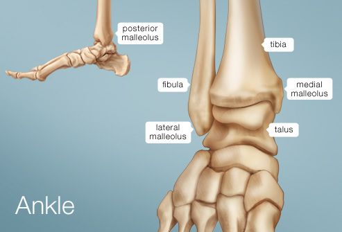
These bones are arranged into two major divisions: The authors explore how digitizing one of the seven basic quality tools—the fishbone diagram—using mind mapping can significantly improve the tool. Related posts of bones leg diagram picture. Image result for leg bones diagram human leg bone structure your leg bones are the longest and strongest bones in your body. Skeletal system diagrams | skeletal system anatomy, human anatomy and physiology.diagram of blood and nerve supply to bone. The tibia, commonly known as the 'shin bone', is the largest and most medial of the two.you can palpate its anterior border when you run your finger down the anterior aspect of your leg. Distal end of right humerus. Click now to learn more about the bones, muscles, and soft tissues tibia: Ulna and the radius are two bones that sit next to each other. Related posts of muscles and tendons of the leg muscle anatomy for gym. Related posts of diagram of leg bones inside of arm muscle and bone. 1 and the one behind the little toe is no. Anatomically, the term leg means the part of the hind limb that extends from the stiffle joint to the hock joint (knee to ankle or tibia and fibula bones region).
The knee joint is the largest joint in the body and is primarily a hinge joint, although some sliding and rotation occur. Muscle anatomy for gym 12 photos of the muscle anatomy for gym muscle anatomy and fitness, muscle anatomy for fitness, muscle anatomy for gym, human muscles, muscle anatomy and fitness, muscle anatomy for fitness, muscle anatomy for gym. Editor · aug 13, 2017 ·. It is made of the ulna and the radius. This short post will try to cover the dog leg anatomy in detail with labeled diagrams.
Pajtu7s1fn6b0m from img.webmd.com 1 and the one behind the little toe is no. He leg's main function in the human is for locomotion and support of the rest of the body. Posted on june 4, 2014 by admin. In fact nearly one quarter of the bones in the body are found in the feet. Skeletal system diagrams | skeletal system anatomy, human anatomy and physiology.diagram of blood and nerve supply to bone. The major bones of the leg are the femur (thigh bone), tibia (shin bone), and adjacent fibula, and these are all long bones. Muscle anatomy for gym 12 photos of the muscle anatomy for gym muscle anatomy and fitness, muscle anatomy for fitness, muscle anatomy for gym, human muscles, muscle anatomy and fitness, muscle anatomy for fitness, muscle anatomy for gym. The femur, or thighbone, is the longest and largest bone in the human body.
The authors explore how digitizing one of the seven basic quality tools—the fishbone diagram—using mind mapping can significantly improve the tool.
1 and the one behind the little toe is no. This diagram depicts diagram leg bones anatomy. The bones together make up the hip. The largest and most medial leg bone, forming both the knee and ankle joints. The hip itself is a ball and socket joint, much like the shoulder.the structures necessary to create this joint are the socket, the joint capsule, muscle, ligaments, and the neck. These muscles work together to produce movements such as standing, walking, running, and jumping. Muscle anatomy for gym 12 photos of the muscle anatomy for gym muscle anatomy and fitness, muscle anatomy for fitness, muscle anatomy for gym, human muscles, muscle anatomy and fitness, muscle anatomy for fitness, muscle anatomy for gym. The tibia and fibula are two long bones that run parallel to each other forming the scaffold of the leg and providing attachment points for many muscles. The elbow is located below the chest at the back of the foreleg. Fish(bone) stories (quality progress) the method behind the fishbone diagram is older than many of its users. The femur, or thighbone, is the longest and largest bone in the human body. The technical term for a dog knee is the stifle joint. Click now to learn more about the bones, muscles, and soft tissues tibia:


0 Komentar